Performance Evaluation of ResNet50 and VGG16 in Automated Multi-Stage Breast Cancer Histopathology Grading
DOI:
https://doi.org/10.12928/biste.v8i5.15824Keywords:
Breast Cancer, Histopathology, Nottingham Histologic Grading, Resnet50, VGG16, Convolutional Neural Network (CNN)Abstract
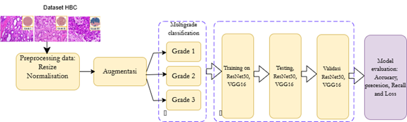
Breast cancer histopathological grading is important for estimating tumor aggressiveness and supporting treatment planning, yet manual assessment can be influenced by observer variability. This study comparatively evaluates two convolutional neural network architectures, ResNet50 and VGG16, for automated multi-stage breast cancer histopathology grading based on the Nottingham histologic grading categories: Grade I, Grade II, and Grade III. The task was formulated as image-level classification using 1,742 hematoxylin and eosin-stained histopathological images obtained from 150 patients at an Anatomical Pathology Laboratory. All images were resized to 224 × 224 pixels, converted into normalized numerical RGB values, and augmented using horizontal and vertical flipping. The dataset was divided into training, validation, and testing subsets while considering patient-level separation to reduce data leakage. VGG16 was trained using stochastic gradient descent with a learning rate of 0.001, a batch size of 32 for training and 64 for testing, and 60 epochs. ResNet50 was trained using stochastic gradient descent and CrossEntropyLoss for 200 epochs, with raw logits in the final layer because CrossEntropyLoss internally applies softmax. Experimental results showed that ResNet50 achieved higher training accuracy (0.9735), higher validation accuracy (0.6792), lower training and validation loss (0.0809), and slightly higher recall (0.7095) than VGG16. Both models obtained the same test accuracy of 0.7590. These findings indicate that ResNet50 demonstrated stronger learning behavior and slightly better sensitivity, although external validation and class-wise analysis remain necessary before clinical application. The study also provides a controlled baseline for future CNN-based decision-support research in digital breast pathology settings and practice.
References
H. Sung, J. Ferlay, R. L. Siegel, M. Laversanne, I. Soerjomataram, A. Jemal, and F. Bray, "Global Cancer Statistics 2020: GLOBOCAN Estimates of Incidence and Mortality Worldwide for 36 Cancers in 185 Countries," CA Cancer J. Clin., vol. 71, no. 3, pp. 209-249, May 2021, https://doi.org/10.3322/caac.21660.
N. Harbeck, F. Penault-Llorca, J. Cortes, M. Gnant, N. Houssami, P. Poortmans, K. Ruddy, E. Tsang, and F. Cardoso, "Breast cancer," Nat. Rev. Dis. Primers, vol. 5, no. 1, p. 66, Sep. 2019, https://doi.org/10.1038/s41572-019-0111-2.
A. G. Waks and E. P. Winer, "Breast Cancer Treatment: A Review," JAMA, vol. 321, no. 3, pp. 288-300, Jan. 2019, https://doi.org/10.1001/jama.2018.19323.
C. van Dooijeweert, P. J. van Diest, and I. O. Ellis, "Grading of invasive breast carcinoma: the way forward," Virchows Arch., vol. 480, no. 1, pp. 33-43, Jan. 2022, https://doi.org/10.1007/s00428-021-03141-2.
P. S. Ginter, R. Idress, T. M. D'Alfonso, S. Fineberg, and M. Harigopal, "Histologic grading of breast carcinoma: a multi-institution study of interobserver variation using virtual microscopy," Mod. Pathol., vol. 34, no. 4, pp. 701-709, Apr. 2021, https://doi.org/10.1038/s41379-020-00698-2.
C. W. Elston and I. O. Ellis, "Pathological prognostic factors in breast cancer. I. The value of histological grade in breast cancer: experience from a large study with long-term follow-up," Histopathology, vol. 19, no. 5, pp. 403-410, Nov. 1991, https://doi.org/10.1111/j.1365-2559.1991.tb00229.x.
E. A. Rakha, M. E. M. El-Sayed, A. R. Lee, I. O. Ellis, and J. F. R. Robertson, "Prognostic significance of Nottingham histologic grade in invasive breast carcinoma," J. Clin. Oncol., vol. 26, no. 19, pp. 3153-3158, Jul. 2008, https://doi.org/10.1200/JCO.2007.15.5986.
E. A. Rakha et al., "Breast cancer prognostic classification in the molecular era: the role of histological grade," Breast Cancer Res., vol. 12, no. 4, p. 207, Aug. 2010, https://doi.org/10.1186/bcr2607.
A. P. Wibawa, A. N. Handayani, M. R. M. Rukantala, M. Ferdyan, L. A. P. Budi, A. B. P. Utama, and F. A. Dwiyanto, "Decoding and preserving Indonesia's iconic Keris via A CNN-based classification," Telematics and Informatics Reports, vol. 13, p. 100120, Mar. 2024, https://doi.org/10.1016/j.teler.2024.100120.
J. van der Laak, G. Litjens, and F. Ciompi, "Deep learning in histopathology: the path to the clinic," Nat. Med., vol. 27, no. 5, pp. 775-784, May 2021, https://doi.org/10.1038/s41591-021-01343-4.
G. Litjens et al., "A survey on deep learning in medical image analysis," Med. Image Anal., vol. 42, pp. 60-88, Dec. 2017, https://doi.org/10.1016/j.media.2017.07.005.
K. He, X. Zhang, S. Ren, and J. Sun, "Deep Residual Learning for Image Recognition," in Proc. IEEE Conf. Comput. Vis. Pattern Recognit. (CVPR), 2016, pp. 770-778, https://doi.org/10.1109/CVPR.2016.90.
K. Simonyan and A. Zisserman, "Very Deep Convolutional Networks for Large-Scale Image Recognition," 2014, arXiv:1409.1556.
H. D. Couture et al., "Image analysis with deep learning to predict breast cancer grade, ER status, histologic subtype, and intrinsic subtype," npj Breast Cancer, vol. 4, p. 30, 2018, https://doi.org/10.1038/s41523-018-0079-1.
Y. Wang et al., "Improved breast cancer histological grading using deep learning," Ann. Oncol., vol. 33, no. 1, pp. 89-98, Jan. 2022, https://doi.org/10.1016/j.annonc.2021.09.007.
R. Jaroensri et al., "Deep learning models for histologic grading of breast cancer and association with disease prognosis," npj Breast Cancer, vol. 8, p. 113, Oct. 2022, https://doi.org/10.1038/s41523-022-00478-y.
L. Jiang et al., "Deep learning applications in breast cancer histopathological imaging: diagnosis, treatment, and prognosis," Breast Cancer Res., vol. 26, p. 95, 2024, https://doi.org/10.1186/s13058-024-01895-6.
M. Gadermayr and M. Tschuchnig, "Multiple instance learning for digital pathology: A review of the state-of-the-art, limitations & future potential," Comput. Med. Imaging Graph., vol. 112, p. 102337, Mar. 2024, https://doi.org/10.1016/j.compmedimag.2024.102337.
G. Campanella et al., "Clinical-grade computational pathology using weakly supervised deep learning on whole slide images," Nat. Med., vol. 25, no. 8, pp. 1301-1309, Aug. 2019, https://doi.org/10.1038/s41591-019-0508-1.
M. Ilse, J. M. Tomczak, and M. Welling, "Attention-based Deep Multiple Instance Learning," in Proc. Int. Conf. Mach. Learn. (ICML), pp. 2127-2136, 2018, https://proceedings.mlr.press/v80/ilse18a.html?ref=https://.
D. Tellez et al., "Neural Image Compression for Gigapixel Histopathology Image Analysis," IEEE Trans. Pattern Anal. Mach. Intell., vol. 43, no. 2, pp. 567-578, Feb. 2021, https://doi.org/10.1109/TPAMI.2019.2936841.
M. I. Mahmud, M. Mamun and A. Abdelgawad, "A Deep Analysis of Transfer Learning Based Breast Cancer Detection Using Histopathology Images," 2023 10th International Conference on Signal Processing and Integrated Networks (SPIN), pp. 198-204, 2023, https://doi.org/10.1109/SPIN57001.2023.10117110.
F. A. Spanhol, L. S. Oliveira, C. Petitjean, and L. Heutte, "A dataset for breast cancer histopathological image classification," IEEE Trans. Biomed. Eng., vol. 63, no. 7, pp. 1455-1462, Jul. 2016, https://doi.org/10.1109/TBME.2015.2496264.
N. Islam, K. M. Hasib, M. F. Mridha, S. Alfarhood, M. Safran, and M. K. Bhuyan, "Fusing global context with multiscale context for enhanced breast cancer classification," Sci. Rep., vol. 14, p. 27358, Nov. 2024, https://doi.org/10.1038/s41598-024-78363-w.
G. Litjens, T. Kooi, B. E. Bejnordi, A. A. A. Setio, F. Ciompi, M. Ghafoorian, J. A. W. M. van der Laak, B. van Ginneken, and C. I. Sánchez, "A survey on deep learning in medical image analysis," Med. Image Anal., vol. 42, pp. 60-88, Dec. 2017, https://doi.org/10.1016/j.media.2017.07.005.
M. Z. Hoque, A. Keskinarkaus, P. Nyberg, and T. Seppänen, "Stain normalization methods for histopathology image analysis: A comprehensive review and experimental comparison," Inf. Fusion, vol. 102, p. 101997, Feb. 2024, https://doi.org/10.1016/j.inffus.2023.101997.
P. A. Dunn, S. Stallard, L. A. E. van der Maaten, S. L. van der Laak, D. J. B. S. Weijers, R. van Dijk, and J. van der Laak, "Stain variability negatively impacts deep learning model generalization in histopathology: An international multi-center study," J. Pathol. Inform., vol. 16, p. 101011, 2025, https://doi.org/10.1016/j.jpi.2025.101011.
M. A. Morid, A. Borjali, and G. Del Fiol, "A scoping review of transfer learning research on medical image analysis using ImageNet," Comput. Biol. Med., vol. 128, p. 104115, Jan. 2021, https://doi.org/10.1016/j.compbiomed.2020.104115.
O. Russakovsky, J. Deng, H. Su, J. Krause, S. Satheesh, S. Ma, Z. Huang, A. Karpathy, A. Khosla, M. Bernstein, A. C. Berg, and L. Fei-Fei, "ImageNet Large Scale Visual Recognition Challenge," Int. J. Comput. Vis., vol. 115, pp. 211-252, 2015, https://doi.org/10.1007/s11263-015-0816-y.
G. Litjens et al., "A survey on deep learning in medical image analysis," Med. Image Anal., vol. 42, pp. 60-88, 2017, https://doi.org/10.1016/j.media.2017.07.005.
R. Sutjiadi, S. Sendari, H. W. Herwanto, and Y. Kristian, "Generating High-quality Synthetic Mammogram Images Using Denoising Diffusion Probabilistic Models: a Novel Approach for Augmenting Deep Learning Datasets," in Proc. 2024 Int. Conf. Information Technology Systems and Innovation (ICITSI), pp. 386-392, Dec. 2024, https://doi.org/10.1109/ICITSI65188.2024.10929446.
R. Sutjiadi, S. Sendari, H. W. Herwanto, and Y. Kristian, "Deep Learning for Segmentation and Classification in Mammograms for Breast Cancer Detection: A Systematic Literature Review," Adv. Ultrasound Diagn. Ther., vol. 8, no. 3, pp. 94-105, 2024, https://doi.org/10.37015/AUDT.2024.230051.
R. Sutjiadi, S. Sendari, H. W. Herwanto, and Y. Kristian, "Leveraging Synthetic Mammograms to Enhance Deep-Learning Performance for Breast Cancer Classification Using EfficientNetV2L Architecture," EAI Endorsed Trans. AI Robot., vol. 4, Sep. 2025, https://doi.org/10.4108/airo.9749.
H. W. Herwanto, "Penggunaan wavelet image enhancement dan tekstur energi citra untuk mendeteksi massa mencurigakan pada mamogram," Teknologi dan Kejuruan, vol. 31, no. 1, Sep. 2012, https://doi.org/10.17977/tk.v31i1.3187.
I. K. M. Jais and A. R. Ismail, "Adam Optimization Algorithm for Wide and Deep Neural Network," Knowledge Engineering and Data Science, vol. 2, no. 1, pp. 41-46, 2019, https://doi.org/10.17977/um018v2i12019p41-46.
F. Schwarzhans et al., "Image normalization techniques and their effect on the robustness and predictive power of breast MRI radiomics," Eur. J. Radiol., vol. 187, p. 112086, 2025, https://doi.org/10.1016/j.ejrad.2025.112086.
M. J. Willemink et al., "Preparing medical imaging data for machine learning," Radiology, vol. 295, no. 1, pp. 4-15, 2020, https://doi.org/10.1148/radiol.2020192224.
C. Shorten and T. M. Khoshgoftaar, "A survey on image data augmentation for deep learning," J. Big Data, vol. 6, p. 60, 2019, https://doi.org/10.1186/s40537-019-0197-0.
D. Tellez et al., "Quantifying the effects of data augmentation and stain color normalization in convolutional neural networks for computational pathology," Med. Image Anal., vol. 58, p. 101544, 2019, https://doi.org/10.1016/j.media.2019.101544.
N. Bussola, A. Marcolini, V. Maggio, G. Jurman, and C. Furlanello, "AI slipping on tiles: Data leakage in digital pathology," in Pattern Recognition. ICPR International Workshops and Challenges, pp. 167-182, 2021, https://doi.org/10.1007/978-3-030-68763-2_13.
I. E. Tampu, A. Eklund, and N. Haj-Hosseini, "Inflation of test accuracy due to data leakage in deep learning-based classification of OCT images," Sci. Data, vol. 9, p. 580, 2022, https://doi.org/10.1038/s41597-022-01618-6.
H. C. Shin, H. R. Roth, M. Gao, L. Lu, Z. Xu, I. Nogues, J. Yao, D. Mollura, and R. M. Summers, "Deep convolutional neural networks for computer-aided detection: CNN architectures, dataset characteristics and transfer learning," IEEE Trans. Med. Imaging, vol. 35, no. 5, pp. 1285-1298, May 2016, https://doi.org/10.1109/TMI.2016.2528162.
S. J. Pan and Q. Yang, "A survey on transfer learning," IEEE Trans. Knowl. Data Eng., vol. 22, no. 10, pp. 1345-1359, Oct. 2010, https://doi.org/10.1109/TKDE.2009.191.
R. Mormont, P. Geurts, and R. Marée, "Comparison of deep transfer learning strategies for digital pathology," in Proc. IEEE Conf. Comput. Vis. Pattern Recognit. Workshops (CVPRW), Jun. 2018, pp. 2262-2271.
M. Sokolova and G. Lapalme, "A systematic analysis of performance measures for classification tasks," Inf. Process. Manage., vol. 45, no. 4, pp. 427-437, Jul. 2009, https://doi.org/10.1016/j.ipm.2009.03.002.
I. Goodfellow, Y. Bengio, and A. Courville. Deep learning. vol. 1, no. 2, pp. 1-800. Cambridge: MIT press. 2016, https://doi.org/10.4258/hir.2016.22.4.351.
M. Macenko, M. Niethammer, J. S. Marron, D. Borland, J. T. Woosley, X. Guan, C. Schmitt, and N. E. Thomas, "A method for normalizing histology slides for quantitative analysis," in Proc. 2009 IEEE Int. Symp. Biomed. Imaging: From Nano to Macro (ISBI), 2009, pp. 1107-1110, https://doi.org/10.1109/ISBI.2009.5193250.
E. Reinhard, M. Ashikhmin, B. Gooch, and P. Shirley, "Color transfer between images," IEEE Comput. Graph. Appl., vol. 21, no. 5, pp. 34-41, Sep.-Oct. 2001, https://doi.org/10.1109/38.946629.
G. Landini, G. Martinelli, and F. Piccinini, “Colour deconvolution: stain unmixing in histological imaging,” Bioinformatics, vol. 37, no. 10, pp. 1485-1487, 2021, https://doi.org/10.1093/bioinformatics/btaa847.
H. Zhang, M. Cisse, Y. N. Dauphin, and D. Lopez-Paz, "mixup: Beyond empirical risk minimization," arXiv:1710.09412, 2017, https://doi.org/10.48550/arXiv.1710.09412.
S. Yun, D. Han, S. J. Oh, S. Chun, J. Choe, and Y. Yoo, "CutMix: Regularization strategy to train strong classifiers with localizable features," In Proceedings of the IEEE/CVF international conference on computer vision, pp. 6023-6032, 2019, https://openaccess.thecvf.com/content_ICCV_2019/html/Yun_CutMix_Regularization_Strategy_to_Train_Strong_Classifiers_With_Localizable_Features_ICCV_2019_paper.html.
E. D. Cubuk, B. Zoph, J. Shlens, and Q. V. Le, "RandAugment: Practical automated data augmentation with a reduced search space," in Proc. IEEE/CVF Conf. Comput. Vis. Pattern Recognit. Workshops (CVPRW), 2020, arXiv:1909.13719.
J. Cohen, "A coefficient of agreement for nominal scales," Educ. Psychol. Meas., vol. 20, no. 1, pp. 37-46, Apr. 1960, https://doi.org/10.1177/001316446002000104.
T. Fawcett, "An introduction to ROC analysis," Pattern Recognit. Lett., vol. 27, no. 8, pp. 861-874, Jun. 2006, https://doi.org/10.1016/j.patrec.2005.10.010.
R. R. Selvaraju, M. Cogswell, A. Das, R. Vedantam, D. Parikh, and D. Batra, "Grad-CAM: Visual explanations from deep networks via gradient-based localization," Int. J. Comput. Vis., vol. 128, no. 2, pp. 336-359, Feb. 2020, https://doi.org/10.1007/s11263-019-01228-7.
Published
How to Cite
Issue
Section
License
Copyright (c) 2026 Agoes Santika Hyperastuty, Anik Nur Handayani, Heru Wahyu Herwanto, Nor Salwa Damanhuriis

This work is licensed under a Creative Commons Attribution-ShareAlike 4.0 International License.
Authors who publish with this journal agree to the following terms:
- Authors retain copyright and grant the journal right of first publication with the work simultaneously licensed under a Creative Commons Attribution License that allows others to share the work with an acknowledgment of the work's authorship and initial publication in this journal.
- Authors are able to enter into separate, additional contractual arrangements for the non-exclusive distribution of the journal's published version of the work (e.g., post it to an institutional repository or publish it in a book), with an acknowledgment of its initial publication in this journal.
- Authors are permitted and encouraged to post their work online (e.g., in institutional repositories or on their website) prior to and during the submission process, as it can lead to productive exchanges, as well as earlier and greater citation of published work (See The Effect of Open Access).

This journal is licensed under a Creative Commons Attribution-ShareAlike 4.0 International License.