Grid-Aligned Patchification for Deep Learning-Based Macrophage Detection in Unstained Brightfield Haemocytometer Images
DOI:
https://doi.org/10.12928/biste.v8i3.16005Keywords:
Brightfield Microscopy, Haemocytometer, Instance Segmentation, Macrophage, Deep LearningAbstract
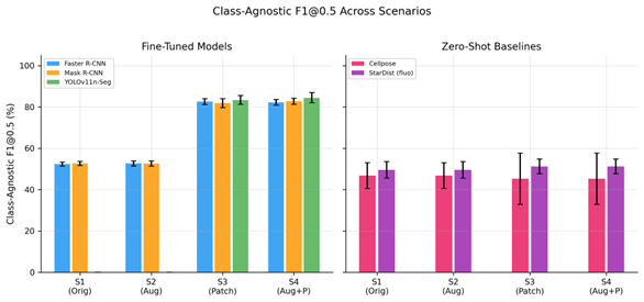
Manual cell counting from haemocytometer images is slow, subjective, and operator-dependent, especially in unstained brightfield microscopy where cell boundaries and viability-related morphology are difficult to distinguish. Although prior cell detection models have mainly been evaluated on stained or fluorescence images, systematic comparisons between fine-tuned detectors and zero-shot cell segmentation models remain limited for unstained brightfield haemocytometer images. This study presents a controlled 2×2 factorial benchmark of patchification and augmentation across five detection approaches, with variance-decomposition analysis and comparison of fine-tuned versus zero-shot deployment modes. Using 24 unstained brightfield RAW 264.7 macrophage images with 6,307 polygon-level annotations, including 28.8% dead cells, we evaluated four preprocessing scenarios under six-fold stratified cross-validation. Faster R-CNN, Mask R-CNN, and YOLOv11n-Seg were fine-tuned within each fold, whereas Cellpose and StarDist were applied zero-shot. Grid-aligned patchification improved bounding-box mAP50 by 2.6–8.4× across all fine-tuned architectures (paired Wilcoxon p = 0.016, Cohen’s d > 3). A 2×2 ANOVA attributed 99.2–99.4% of explained variance to patchification, while augmentation and interaction effects each contributed less than 0.1%, suggesting that performance gains were driven mainly by scale rescaling rather than sample count. On patchified data, fine-tuned models converged to 85.5–86.4% mAP50. YOLOv11n-Seg achieved the highest mAP50-95 of 51.1%, with 6× faster inference and 17× fewer parameters. In contrast, zero-shot Cellpose and StarDist reached only 45.3–51.2% class-agnostic F1@0.5. These findings show that structure-aware patchification is critical for reliable cell detection in this modality.
References
T. L. Riss, R. A. Moravec, A. L. Niles, S. Duellman, H. A. Benink, T. J. Worzella, and L. Minor, “Cell viability assays,” Assay guidance manual [Internet], 2016, https://www.ncbi.nlm.nih.gov/books/NBK574243/?report=reader.
K. A. Patel et al., “Validation of Automated Fluorescent-Based Technology for Measuring Total Nucleated Cell Viability of Hematopoietic Progenitor Cell Products,” Transfusion (Paris), vol. 62, no. 4, pp. 848–856, 2022, https://doi.org/10.1111/trf.16837.
W. Strober, “Trypan Blue Exclusion Test of Cell Viability,” Curr. Protoc. Immunol., vol. 111, no. 1, p. A3.B.1-A3.B.3, 2015, https://doi.org/10.1002/0471142735.ima03bs111.
Y. Chen and P.-J. Chiang, “An Automated Approach for Hemocytometer Cell Counting Based on Image-Processing Method,” Measurement, vol. 234, p. 114894, 2024, https://doi.org/10.1016/j.measurement.2024.114894.
K. Hildebrand et al., “AI-Driven Analysis for Real-Time Detection of Unstained Microscopic Cell Culture Images,” AI, vol. 6, no. 10, p. 271, 2025, https://doi.org/10.3390/ai6100271.
Z. Zou, K. Chen, Z. Shi, Y. Guo, and J. Ye, “Object Detection in 20 Years: A Survey,” Proc. IEEE, vol. 111, no. 3, pp. 257–332, 2023, https://doi.org/10.1109/jproc.2023.3238524.
R. Morelli et al., “Automating Cell Counting in Fluorescent Microscopy through Deep Learning with c-ResUnet,” Sci. Rep., vol. 11, no. 1, 2021, https://doi.org/10.1038/s41598-021-01929-5.
Y. Liu et al., “Cell Counting in the Era of Deep Learning: Methods and Challenges,” Cell Transplant., vol. 33, 2024, https://doi.org/10.1177/09636897241293628.
J. Ma et al., “The Multimodality Cell Segmentation Challenge: Toward Universal Solutions,” Nat. Methods, vol. 21, no. 6, pp. 1103–1113, 2024, https://doi.org/10.1038/s41592-024-02233-6.
S. Ren, K. He, R. Girshick and J. Sun, "Faster R-CNN: Towards Real-Time Object Detection with Region Proposal Networks," in IEEE Transactions on Pattern Analysis and Machine Intelligence, vol. 39, no. 6, pp. 1137-1149, 2017, https://doi.org/10.1109/TPAMI.2016.2577031.
J. Redmon, S. Divvala, R. Girshick, and A. Farhadi, “You Only Look Once: Unified, Real-Time Object Detection,” in Proceedings of the IEEE Conference on Computer Vision and Pattern Recognition, pp. 779–788, 2016, https://doi.org/10.1109/cvpr.2016.91.
J. Terven, D.-M. Cordova-Esparza, and J.-A. Romero-Gonzalez, “A Comprehensive Review of YOLO Architectures in Computer Vision: From YOLOv1 to YOLOv8 and YOLO-NAS,” Mach. Vis. Appl., vol. 34, no. 6, p. 116, 2023, https://doi.org/10.1007/s00138-023-01426-z.
P. Mehta et al., “Benchmarking YOLO Variants for Enhanced Blood Cell Detection,” Int. J. Imaging Syst. Technol., vol. 35, no. 1, 2025, https://doi.org/10.1002/ima.70037.
Y. He, “Automatic Blood Cell Detection Based on Advanced YOLOv5s Network,” IEEE Access, vol. 12, pp. 17639–17650, 2024, https://doi.org/10.1109/access.2024.3360142.
X. Chen et al., “NBCDC-YOLOv8: A new framework to improve blood cell detection and classification based on YOLOv8,” IET Comput. Vis., vol. 19, no. 1, 2025, https://doi.org/10.1049/cvi2.12341.
D. Zhang et al., “TW-YOLO: An Innovative Blood Cell Detection Model Based on Multi-Scale Feature Fusion,” Sensors, vol. 24, no. 19, p. 6168, 2024, https://doi.org/10.3390/s24196168.
M. G. Ragab et al., “A Comprehensive Systematic Review of YOLO for Medical Object Detection (2018 to 2023),” IEEE Access, vol. 12, pp. 57815–57836, 2024, https://doi.org/10.1109/access.2024.3386826.
H. Sazak and M. Kotan, “Automated Blood Cell Detection and Classification in Microscopic Images Using YOLOv11 and Optimized Weights,” Diagnostics, vol. 15, no. 1, p. 22, 2024, https://doi.org/10.3390/diagnostics15010022.
K. He, G. Gkioxari, P. Dollár, and R. Girshick, “Mask R-CNN,” in Proceedings of the IEEE International Conference on Computer Vision, pp. 2961–2969, 2017, https://doi.org/10.1109/ICCV.2017.322.
M. Hu et al., “An Improved Mask R-CNN Model for Cell Detection and Segmentation in Microscopy Images,” Sensors, vol. 24, no. 8, p. 2424, 2024, https://doi.org/10.3390/s24082424.
G. Moallem et al., “Detecting and Counting Unstained and Unmanipulated Adherent Cells in Brightfield by Faster Region-Based Convolutional Neural Network,” J. Biomed. Opt., vol. 27, no. 7, p. 076003, 2022, https://doi.org/10.1117/1.jbo.27.7.076003.
T.-Y. Lin et al., “Microsoft COCO: Common Objects in Context,” in European Conference on Computer Vision, pp. 740–755, 2014, https://doi.org/10.1007/978-3-319-10602-1_48.
K. He, X. Zhang, S. Ren, and J. Sun, “Deep Residual Learning for Image Recognition,” in Proceedings of the IEEE Conference on Computer Vision and Pattern Recognition, pp. 770–778, 2016,https://doi.org/10.1109/cvpr.2016.90.
G. Zhan et al., “Auto-CSC: A Transfer Learning Based Automatic Cell Segmentation and Count Framework,” Cyborg Bionic Syst., vol. 2022, p. 9842349, 2022, https://doi.org/10.34133/2022/9842349.
X. Wang et al., “Induced Pluripotent Stem Cells Detection via Ensemble YOLO Network,” in Proceedings of the Annual International Conference of the IEEE Engineering in Medicine and Biology Society, pp. 3738–3741, 2021, https://doi.org/10.1109/embc46164.2021.9629744.
C. Xu et al., “Transfer Learning and SE-ResNet152 Networks-Based for Small-Scale Unbalanced Cervical Cell Detection,” Diagnostics, vol. 12, no. 10, p. 2477, 2022, https://doi.org/10.3390/diagnostics12102477.
C. Stringer, T. Wang, M. Michaelos, and M. Pachitariu, “Cellpose: a generalist algorithm for cellular segmentation,” Nat. Methods, vol. 18, no. 1, pp. 100–106, 2021, https://doi.org/10.1038/s41592-020-01018-x.
M. Pachitariu and C. Stringer, “Cellpose 2.0: how to train your own model,” Nat. Methods, vol. 19, no. 12, pp. 1634–1641, 2022, https://doi.org/10.1038/s41592-022-01663-4.
U. Schmidt, M. Weigert, C. Broaddus, and G. Myers, “Cell Detection with Star-Convex Polygons,” in Medical Image Computing and Computer Assisted Intervention – MICCAI 2018, pp. 265–273, 2018, https://doi.org/10.1007/978-3-030-00934-2_30.
C. Edlund et al., “LIVECell—A large-scale dataset for label-free live cell segmentation,” Nat. Methods, vol. 18, no. 9, pp. 1038–1045, 2021, https://doi.org/10.1038/s41592-021-01249-6.
L. Kuijpers et al., “Automated cell counting for Trypan blue-stained cell cultures using machine learning,” PLOS ONE, vol. 18, no. 11, p. e0291625, 2023, https://doi.org/10.1371/journal.pone.0291625.
K. Hoyos and W. Hoyos, “Supporting Malaria Diagnosis Using Deep Learning and Data Augmentation,” Diagnostics, vol. 14, no. 7, p. 690, 2024, https://doi.org/10.3390/diagnostics14070690.
D. Fishman et al., “Practical Segmentation of Nuclei in Brightfield Cell Images with Neural Networks Trained on Fluorescently Labelled Samples,” J. Microsc., vol. 284, no. 1, pp. 12–24, 2021, https://doi.org/10.1111/jmi.13038.
M. A. S. Ali et al., “Evaluating Very Deep Convolutional Neural Networks for Nucleus Segmentation from Brightfield Cell Microscopy Images,” SLAS Discov., vol. 26, no. 9, pp. 1125–1137, 2021, https://doi.org/10.1177/24725552211023214.
M. Vašinková et al., “Comparing Deep Learning Performance for Chronic Lymphocytic Leukaemia Cell Segmentation in Brightfield Microscopy Images,” Bioinforma. Biol. Insights, vol. 18, 2024, https://doi.org/10.1177/11779322241272387.
S. Korkut, C. Erkan, and S. Aksoy, “On the benefits of region of interest detection for whole slide image classification,” in Proceedings of SPIE, 2023. https://doi.org/10.1117/12.2654193.
D. K. Ufuktepe et al., “Cell Patch Extraction for Malaria Detection Using Deep Learning,” in IEEE Applied Imagery Pattern Recognition Workshop, 2021. https://doi.org/10.1109/aipr52630.2021.9762109.
F. C. Akyon, S. O. Altinuc, and A. Temizel, “Slicing Aided Hyper Inference and Fine-Tuning for Small Object Detection,” in IEEE International Conference on Image Processing, pp. 966–970, 2022,https://doi.org/10.1109/icip46576.2022.9897990.
F. O. Unel, B. O. Ozkalayci, and C. Cigla, “The Power of Tiling for Small Object Detection,” in 2019 IEEE/CVF Conference on Computer Vision and Pattern Recognition Workshops (CVPRW), Long Beach, CA, USA: IEEE, Jun. pp. 582–591, 2019, https://doi.org/10.1109/CVPRW.2019.00084.
S. Das, G. Roy, and P. Zun, “High-Throughput Low-Cost Segmentation of Brightfield Microscopy Live Cell Images,” arXiv preprint arXiv:2508.14106, 2025, https://doi.org/10.48550/arXiv.2508.14106.
G. Moallem, A. A. Pore, A. Gangadhar, H. Sari-Sarraf, and S. A. Vanapalli, “Detection of live breast cancer cells in bright-field microscopy images containing white blood cells by image analysis and deep learning,” J. Biomed. Opt., vol. 27, no. 07, 2022, https://doi.org/10.1117/1.JBO.27.7.076003.
H. Li et al., “Machine-Learning-Assisted Automatic Counting of Fungal Cells in a Hemocytometer,” Eng. Life Sci., vol. 21, no. 12, pp. 835–844, 2021, https://doi.org/10.1002/elsc.202100055.
S. Nakarmi et al., “Deep-Learning Assisted Detection and Quantification of (oo)Cysts of Giardia and Cryptosporidium on Smartphone Microscopy Images,” J. Mach. Learn. Biomed. Imaging, vol. 2, pp. 956–976, 2024, https://doi.org/10.59275/j.melba.2024-a333.
A. Kirillov et al., Segment anything. In Proceedings of the IEEE/CVF international conference on computer vision, pp. 4015-4026, 2023, https://doi.org/10.1109/ICCV51070.2023.00371.
A. Kos, D. Belter, and K. Majek, “Deep Learning for Small and Tiny Object Detection: A Survey,” Pomiary Autom. Robot., vol. 27, no. 3, pp. 85–94, 2023, https://doi.org/10.14313/par_249/85.
F. A. Shewajo and K. A. Fante, “Tile-based microscopic image processing for malaria screening using a deep learning approach,” BMC Med. Imaging, vol. 23, no. 1, p. 39, 2023, https://doi.org/10.1186/s12880-023-00993-9.
F. Abdurahman, K. A. Fante, and M. Aliy, “Malaria parasite detection in thick blood smear microscopic images using modified YOLOV3 and YOLOV4 models,” BMC Bioinformatics, vol. 22, no. 1, p. 112, 2021, https://doi.org/10.1186/s12859-021-04036-4.
T.-Y. Lin, P. Dollár, R. Girshick, K. He, B. Hariharan, and S. Belongie, “Feature Pyramid Networks for Object Detection,” in Proceedings of the IEEE Conference on Computer Vision and Pattern Recognition, pp. 2117–2125, 2017, https://doi.org/10.1109/cvpr.2017.106.
N. E. M. Khalifa, M. Loey, and S. Mirjalili, “A Comprehensive Survey of Recent Trends in Deep Learning for Digital Images Augmentation,” Artif. Intell. Rev., vol. 55, no. 3, pp. 2351–2377, 2022, https://doi.org/10.1007/s10462-021-10066-4.
P. Chlap et al., “A Review of Medical Image Data Augmentation Techniques for Deep Learning Applications,” J. Med. Imaging Radiat. Oncol., vol. 65, no. 5, pp. 545–563, 2021, https://doi.org/10.1111/1754-9485.13261.
A. Kebaili, J. Lapuyade-Lahorgue, and S. Ruan, “Deep Learning Approaches for Data Augmentation in Medical Imaging: A Review,” J. Imaging, vol. 9, no. 4, p. 81, 2023, https://doi.org/10.3390/jimaging9040081.
R. Sapkota and M. Karkee, “Ultralytics YOLO evolution: An overview of YOLO26, YOLO11, YOLOv8 and YOLOv5 object detectors for computer vision and pattern recognition,” arXiv preprint arXiv:2510.09653, 2025, https://doi.org/10.48550/arXiv.2510.09653.
R. Khanam and M. Hussain, “YOLOv11: An Overview of the Key Architectural Enhancements,” arXiv:2410.17725, 2024, https://doi.org/10.48550/arXiv.2410.17725.
Published
How to Cite
Issue
Section
License
Copyright (c) 2026 Mohammad Ikhsan, Zino Ramdani Suharto, Rizal Azis, Basari Basari

This work is licensed under a Creative Commons Attribution-ShareAlike 4.0 International License.
Authors who publish with this journal agree to the following terms:
- Authors retain copyright and grant the journal right of first publication with the work simultaneously licensed under a Creative Commons Attribution License that allows others to share the work with an acknowledgment of the work's authorship and initial publication in this journal.
- Authors are able to enter into separate, additional contractual arrangements for the non-exclusive distribution of the journal's published version of the work (e.g., post it to an institutional repository or publish it in a book), with an acknowledgment of its initial publication in this journal.
- Authors are permitted and encouraged to post their work online (e.g., in institutional repositories or on their website) prior to and during the submission process, as it can lead to productive exchanges, as well as earlier and greater citation of published work (See The Effect of Open Access).

This journal is licensed under a Creative Commons Attribution-ShareAlike 4.0 International License.