CNN-Based Transfer Learning Models for Histopathological Detection of Non-Hodgkin Lymphoma on Histopathological Images
DOI:
https://doi.org/10.12928/biste.v8i2.15207Keywords:
Classification, CNN, Histopathology, Lymphoma, Transfer LearningAbstract
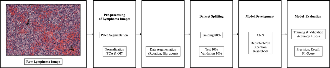
More than 85.720 new cases and 21.000 fatalities from lymphoma were reported globally in 2021. This type of cancer can spread through the body using the lymphatic system and then enter the blood. Since lymphoma affects the lymphatic system, it can be hard to diagnose correctly because there are many different subtypes, such as Mantle Cell Lymphoma (MCL), Follicular Lymphoma (FL), and Chronic Lymphocytic Leukemia (CLL). The diagnostic complexity of lymphoma highlights the need for more accurate and reliable automated diagnostic methods. This research proposes a transfer learning approach employing pre-trained Convolutional Neural Network (CNN) models using DenseNet-201, Xception, and ResNet-50, for lymphoma subtype classification. The dataset consists of microscopic histopathology images from three lymphoma classes (MCL, FL, and CLL). Each image was resized and segmented into 24 non-overlapping patches, followed by Macenko stain normalization and data augmentation. Model performance was evaluated using a random sampling with a fixed random seed train–validation–test split, and validated using cross-validation method. The proposed approach achieved classification accuracies of 96.7% for DenseNet-201, 97.15% for Xception, and 96.3% for ResNet-50. These results indicate that deeper architectures with efficient feature reuse and depthwise separable convolutions improve the detection of subtle morphological differences among lymphoma subtypes. Despite limitations related to dataset size and external validation, the findings demonstrate the potential of transfer learning-based CNN models as decision-support tools for lymphoma diagnosis.
References
S. Soltane, S. Alsharif, and S. M. Serag Eldin, “Classification and diagnosis of lymphoma’s histopathological images using transfer learning,” Computer Systems Science and Engineering, vol. 40, no. 2, pp. 629–644, 2021, https://doi.org/10.32604/CSSE.2022.019333.
V. Gaidano et al., “A clinically applicable approach to the classification of B-cell non-hodgkin lymphomas with flow cytometry and machine learning,” Cancers (Basel), vol. 12, no. 6, pp. 1–18, 2020, https://doi.org/10.3390/cancers12061684.
J. Carreras, N. Nakamura, and R. Hamoudi, “Artificial Intelligence Analysis of Gene Expression Predicted the Overall Survival of Mantle Cell Lymphoma and a Large Pan-Cancer Series,” Healthcare (Switzerland), vol. 10, no. 1, 2022, https://doi.org/10.3390/healthcare10010155.
H. El Achi and J. D. Khoury, “Artificial intelligence and digital microscopy applications in diagnostic hematopathology,” Cancers (Basel), vol. 12, no. 4, pp. 1–15, 2020, https://doi.org/10.3390/cancers12040797.
Z. Swiderska-Chadaj, K. M. Hebeda, M. van den Brand, and G. Litjens, “Artificial intelligence to detect MYC translocation in slides of diffuse large B-cell lymphoma,” Virchows Archiv, vol. 479, no. 3, pp. 617–621, 2021, https://doi.org/10.1007/s00428-020-02931-4.
H. Miyoshi et al., “Deep learning shows the capability of high-level computer-aided diagnosis in malignant lymphoma,” Laboratory Investigation, vol. 100, no. 10, pp. 1300–1310, 2020, https://doi.org/10.1038/s41374-020-0442-3.
K. Hübel, “Lymphoma: New Diagnosis and Current Treatment Strategies,” J Clin Med, vol. 11, no. 6, pp. 10–12, 2022, https://doi.org/10.3390/jcm11061701.
W. Liu et al., “Improving survival of 3760 patients with lymphoma: Experience of an academic center over two decades,” Cancer Med, vol. 9, no. 11, pp. 3765–3774, 2020, https://doi.org/10.1002/cam4.3037.
T. Doeleman, L. M. Hondelink, M. H. Vermeer, M. R. van Dijk, and A. M. R. Schrader, “Artificial intelligence in digital pathology of cutaneous lymphomas: A review of the current state and future perspectives,” Semin Cancer Biol, vol. 94, no. June, pp. 81–88, 2023, https://doi.org/10.1016/j.semcancer.2023.06.004.
K. Thomsen, A. L. Christensen, L. Iversen, H. B. Lomholt, and O. Winther, “Deep Learning for Diagnostic Binary Classification of Multiple-Lesion Skin Diseases,” Front Med (Lausanne), vol. 7, pp. 1–7, 2020, https://doi.org/10.3389/fmed.2020.574329.
W. D. Lewis, S. Lilly, and K. L. Jones, “Lymphoma: Diagnosis and treatment,” American Academy of Family Physicians, vol. 101, no. 1, pp. 34-41, 2020, https://www.aafp.org/pubs/afp/issues/2020/0101/p34.html.
H. Wang, Z. Liu, J. Yang, L. Sheng, and D. Chen, “Using Machine Learning to Expand the Ann Arbor Staging System for Hodgkin and Non-Hodgkin Lymphoma,” BioMedInformatics, vol. 3, no. 3, pp. 514–525, 2023, https://doi.org/10.3390/biomedinformatics3030035.
G. Steinbuss et al., “Deep learning for the classification of non-hodgkin lymphoma on histopathological images,” Cancers (Basel), vol. 13, no. 10, pp. 1–11, 2021, https://doi.org/10.3390/cancers13102419.
M. Hamdi, E. M. Senan, M. E. Jadhav, F. Olayah, B. Awaji, and K. M. Alalayah, “Hybrid Models Based on Fusion Features of a CNN and Handcrafted Features for Accurate Histopathological Image Analysis for Diagnosing Malignant Lymphomas,” Diagnostics, vol. 13, no. 13, 2023, https://doi.org/10.3390/diagnostics13132258.
R. Ferjaoui, M. A. Cherni, S. Boujnah, N. E. H. Kraiem, and T. Kraiem, “Machine learning for evolutive lymphoma and residual masses recognition in whole body diffusion weighted magnetic resonance images,” Comput Methods Programs Biomed, vol. 209, p. 106320, 2021, https://doi.org/10.1016/j.cmpb.2021.106320.
M. R. Reena and P. M. Ameer, “A content-based image retrieval system for the diagnosis of lymphoma using blood micrographs: An incorporation of deep learning with a traditional learning approach,” Comput Biol Med, vol. 145, p. 105463, 2022, https://doi.org/10.1016/j.compbiomed.2022.105463.
C. Xu et al., “A hybrid few-shot multiple-instance learning model predicting the aggressiveness of lymphoma in PET/CT images,” Comput Methods Programs Biomed, vol. 243, p. 107872, 2024, https://doi.org/10.1016/j.cmpb.2023.107872.
S. Basu, R. Agarwal, and V. Srivastava, “Deep discriminative learning model with calibrated attention map for the automated diagnosis of diffuse large B-cell lymphoma,” Biomed Signal Process Control, vol. 76, p. 103728, 2022, https://doi.org/10.1016/j.bspc.2022.103728.
N. Brancati, G. D. Pietro, M. Frucci, and D. Riccio, “A deep learning approach for breast invasive ductal carcinoma detection and lymphoma multi-classification in histological images,” IEEE, vol.7, 2019, https://doi.org/10.1109/ACCESS.2019.2908724.
Y. Guo, P. Decazes, S. Becker, H. Li, and S. Ruan, “Deep Disentangled Representation Learning of Pet Images for Lymphoma Outcome Prediction,” Proceedings - International Symposium on Biomedical Imaging, pp. 2018–2021, 2020, https://doi.org/10.1109/ISBI45749.2020.9098477.
D. Li et al., “A deep learning diagnostic platform for diffuse large B-cell lymphoma with high accuracy across multiple hospitals,” Nat Commun, vol. 11, no. 1, 2020, https://doi.org/10.1038/s41467-020-19817-3.
C. Syrykh et al., “Accurate diagnosis of lymphoma on whole-slide histopathology images using deep learning,” NPJ digital medicine, vol. 3, no. 1, p. 63, 2020, https://doi.org/10.1038/s41746-020-0272-0.
U. V. Somaratne, K. W. Wong, J. Parry, F. Sohel, X. Wang and H. Laga, "Improving Follicular Lymphoma Identification using the Class of Interest for Transfer Learning," 2019 Digital Image Computing: Techniques and Applications (DICTA), pp. 1-7, 2019, https://doi.org/10.1109/DICTA47822.2019.8946075.
X. Zhang, K. Zhang, M. Jiang, and L. Yang, “Research on the classification of lymphoma pathological images based on deep residual neural network,” Technology and Health Care, vol. 29, pp. S335–S344, 2021, https://doi.org/10.3233/THC-218031.
B. Sheng, M. Zhou, M. Hu, Q. Li, L. Sun, and Y. Wen, “A blood cell dataset for lymphoma classification using faster R-CNN,” Biotechnology and Biotechnological Equipment, vol. 34, no. 1, pp. 413–420, 2020, https://doi.org/10.1080/13102818.2020.1765871.
I. Häggström et al., “Deep learning for [18F]fluorodeoxyglucose-PET-CT classification in patients with lymphoma: a dual-centre retrospective analysis,” Lancet Digit Health, vol. 6, no. 2, pp. e114–e125, 2024, https://doi.org/10.1016/S2589-7500(23)00203-0.
R. Tambe, S. Mahajan, U. Shah, M. Agrawal, and B. Garware, “Towards designing an automated classification of lymphoma subtypes using deep neural networks,” ACM International Conference Proceeding Series, pp. 143–149, 2019, https://doi.org/10.1145/3297001.3297019.
S. Rajadurai, K. Perumal, M. F. Ijaz, and C. L. Chowdhary, “PrecisionLymphoNet: advancing malignant lymphoma diagnosis via ensemble transfer learning with CNNs,” Diagnostics, vol. 14, no. 5, p. 469, 2024, https://doi.org/10.3390/diagnostics14050469.
J. Zhang, W. Cui, X. Guo, B. Wang, and Z. Wang, “Classification of digital pathological images of non-Hodgkin’s lymphoma subtypes based on the fusion of transfer learning and principal component analysis,” Med Phys, vol. 47, no. 9, pp. 4241–4253, 2020, https://doi.org/10.1002/mp.14357.
A. Haghofer et al., “Histological classification of canine and feline lymphoma using a modular approach based on deep learning and advanced image processing,” Sci Rep, vol. 13, no. 1, pp. 1–15, 2023, https://doi.org/10.1038/s41598-023-46607-w.
M. U. Nasir et al., “Hematologic Cancer Detection Using White Blood Cancerous Cells Empowered with Transfer Learning and Image Processing,” J Healthc Eng, vol. 2023, no. 1, p. 1406545 2023, https://doi.org/10.1155/2023/1406545.
N. Kaur and A. Singh, "A Novel Advanced Transfer Learning Based Approach for Leukemia Detection," 2024 International Conference on Cybernation and Computation (CYBERCOM), pp. 415-419, 2024, https://doi.org/10.1109/CYBERCOM63683.2024.10803147.
M. A. Elaziz et al., “Medical image classification utilizing ensemble learning and Levy flight-based honey badger algorithm on 6G-enabled Internet of Things,” Computational Intelligence and Neuroscience, vol. 2022, no. 1, p. 5830766, 2022, https://doi.org/10.1155/2022/5830766.
I. N. Muhsen, D. Shyr, A. D. Sung, and S. K. Hashmi, “Machine Learning Applications in the Diagnosis of Benign and Malignant Hematological Diseases,” Clin Hematol Int, vol. 3, no. 1, p. 13, 2020, https://doi.org/10.2991/chi.k.201130.001.
L. Irshaid et al., “Histopathologic and Machine Deep Learning Criteria to Predict Lymphoma Transformation in Bone Marrow Biopsies,” Arch Pathol Lab Med, vol. 146, no. 2, pp. 182–193, 2022, https://doi.org/10.5858/ARPA.2020-0510-OA.
C. S. Lisson et al., “Deep Neural Networks and Machine Learning Radiomics Modelling for Prediction of Relapse in Mantle Cell Lymphoma,” Cancers (Basel), vol. 14, no. 8, 2022, https://doi.org/10.3390/cancers14082008.
J. Uddin, “Attention-Based DenseNet for Lung Cancer Classification Using CT Scan and Histopathological Images,” Designs (Basel), vol. 8, no. 2, 2024, https://doi.org/10.3390/designs8020027.
Z. Zhong, M. Zheng, H. Mai, J. Zhao, and X. Liu, “Cancer image classification based on DenseNet model,” J Phys Conf Ser, vol. 1651, no. 1, 2020, https://doi.org/10.1088/1742-6596/1651/1/012143.
J. S. Mohlman, S. D. Leventhal, T. Hansen, J. Kohan, V. Pascucci, and M. E. Salama, “Improving Augmented Human Intelligence to Distinguish Burkitt Lymphoma from Diffuse Large B-Cell Lymphoma Cases,” Am J Clin Pathol, vol. 153, no. 6, pp. 743–759, 2020, https://doi.org/10.1093/AJCP/AQAA001.
C. Perry et al., “Image-Based Deep Learning Detection of High-Grade B-Cell Lymphomas Directly from Hematoxylin and Eosin Images,” Cancers (Basel), vol. 15, no. 21, 2023, https://doi.org/10.3390/cancers15215205.
H. E. Achi et al., “Automated diagnosis of lymphoma with digital pathology images using deep learning,” Annals of Clinical & Laboratory Science, vol.49, no.2, 2019, https://www.annclinlabsci.org/content/49/2/153.short.
Published
How to Cite
Issue
Section
License
Copyright (c) 2026 Aghnia Hasya Affan, Basari Basari

This work is licensed under a Creative Commons Attribution-ShareAlike 4.0 International License.
Authors who publish with this journal agree to the following terms:
- Authors retain copyright and grant the journal right of first publication with the work simultaneously licensed under a Creative Commons Attribution License that allows others to share the work with an acknowledgment of the work's authorship and initial publication in this journal.
- Authors are able to enter into separate, additional contractual arrangements for the non-exclusive distribution of the journal's published version of the work (e.g., post it to an institutional repository or publish it in a book), with an acknowledgment of its initial publication in this journal.
- Authors are permitted and encouraged to post their work online (e.g., in institutional repositories or on their website) prior to and during the submission process, as it can lead to productive exchanges, as well as earlier and greater citation of published work (See The Effect of Open Access).

This journal is licensed under a Creative Commons Attribution-ShareAlike 4.0 International License.