CNN Classification of Malaria Parasites in Digital Microscope Images Using Python on Raspberry Pi
DOI:
https://doi.org/10.12928/biste.v5i1.7522Keywords:
Malaria Parasites, Microscope Image, Raspberry Pi Minicomputer, CNN Classification, PythonsAbstract
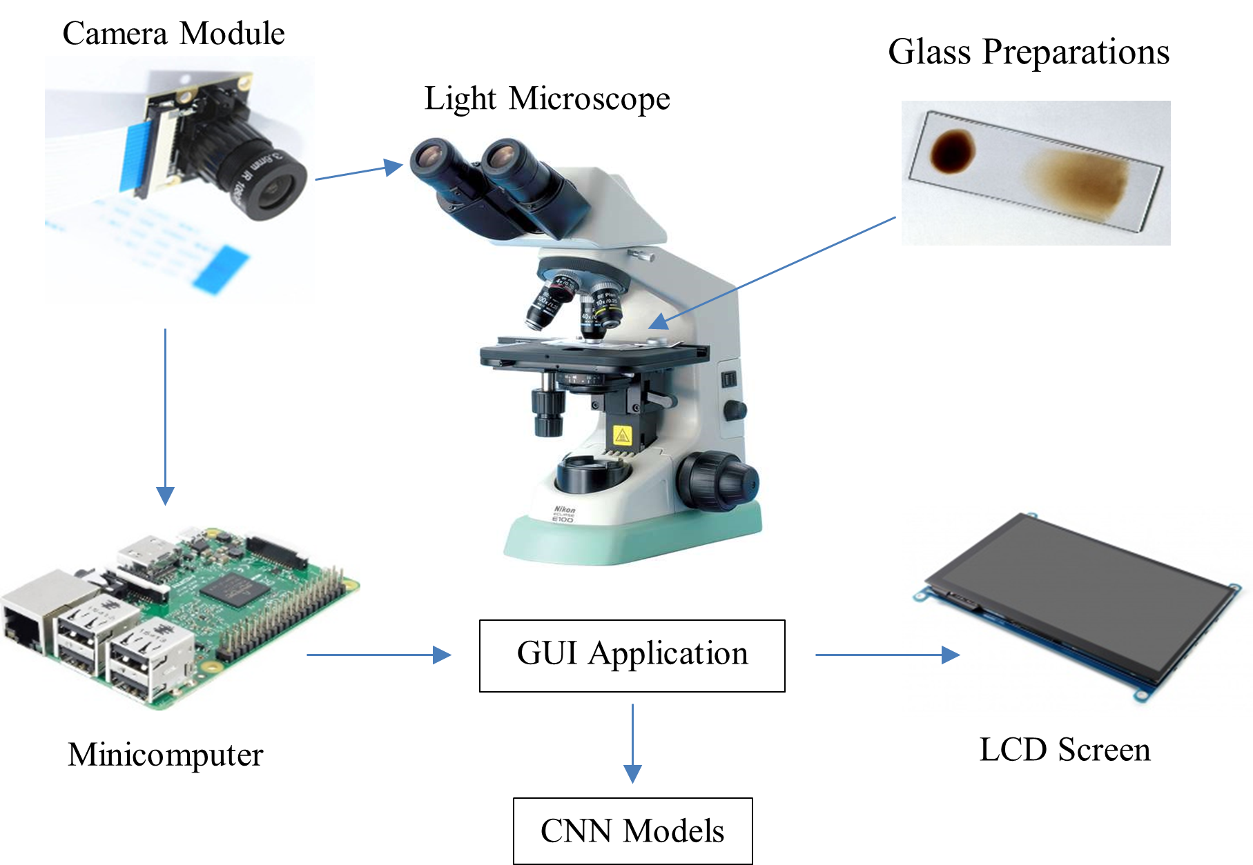
Malaria disease occurs because the plasmodium parasite infects human red blood cells spread by female Anopheles mosquitoes and then causes health problems such as blood deficiency and even death. The gold standard of malaria diagnosis is to use laboratory microscopy examination of the patient's red blood cell samples to distinguish between microscope images of parasitic and non-parasitic red blood cells. However, diagnosing malaria through microscope observation is time-consuming, subjective, and tiring for health workers. So, a malaria classification system was designed using the Convolutional Neural Network (CNN) method to distinguish parasitic and non-parasitic red blood cell images. The CNN model is trained using training data and also tested using test data. Then, the CNN training model is embedded on a Raspberry Pi equipped with a Graphical User Interface to facilitate observer interaction through the LCD screen on this digital and portable microscope. The CNN classification rate achieved an accuracy value of 97.88% using the database image and 98.76% using the digital microscope acquisition image. The CNN classification system of malaria parasites designed on a Raspberry Pi-based digital and portable microscope is expected to improve the diagnosis of malaria and reduce the infection rate of malaria patients, especially in various remote areas in Indonesia.
References
E. L. Febrianti and T. Christy, “Penerapan Forward Chaining Untuk Mendiagnosa Penyakit Malaria dan Pencegahannya Berbasis Web,” Jurnal Teknologi dan Sistem Informasi, vol. 4, no. 1, pp. 93-100, 2017, https://doi.org/10.33330/jurteksi.v4i1.32.
N. K. C. Pratiwi, N. Ibrahim, Y. N. Fu’adah and S. Rizal, “Deteksi Parasit Plasmodium pada Citra Mikroskopis Hapusan Darah dengan Metode Deep Learning,” ELKOMIKA: Jurnal Teknik Energi Elektrik, Teknik Telekomunikasi, & Teknik Elektronika, vol. 9, no. 2, pp. 306-317, 2021, https://doi.org/10.26760/elkomika.v9i2.306.
Direktorat Jenderal Pencegahan dan Pengendalian Penyakit, Kemenkes RI, “Buku Saku Tata Laksana Kasus Malaria.” 2020, https://persi.or.id/wp-content/uploads/2020/11/bukusaku_malaria.pdf.
A. W. Setiawan, A. Faisal, N. Resfita and Y. A. Rahman, “Detection of Malaria Parasites using Thresholding in RGB, YCbCr and Lab Color Spaces,” 2021 International Seminar on Application for Technology of Information and Communication (iSemantic), pp. 70-75, 2021, https://doi.org/10.1109/iSemantic52711.2021.9573224.
Yohannes, S. Devella and K. Arianto, “Deteksi Penyakit Malaria Menggunakan Convolutional Neural Network Berbasis Saliency,” JUITA: Jurnal Informatika, vol. 8, no. 1, pp. 37–44, 2020, https://doi.org/10.30595/juita.v8i1.6671.
K. I. Djahari and G. Hermawan, “Implementasi Metode Principal Component Analysis dan Support Vector Machines dalam Mendeteksi Plasmodium Malaria pada Citra Sampel Darah,” Teknik Informatika – Universitas Komputer Indonesia, 2016, http://repository.unikom.ac.id/id/eprint/53445.
M. Hamid, P. Mudjirahardjo and E. Yudaningtyas, “Penerapan Fitur Warna Untuk Identifikasi Plasmodium Falciparum pada Sediaan Apus Darah Menggunakan MK-Means dan Jaringan Backpropagation,” MATICS: Jurnal Ilmu Komputer dan Teknologi Informasi, vol. 8, no. 2, pp. 73–77, 2016, https://doi.org/10.18860/mat.v8i2.3730.
M. O. Arowolo, M. Adebiyi, A. Adebiyi and O. Okesola, “PCA Model For RNA-Seq Malaria Vector Data Classification Using KNN And Decision Tree Algorithm,” 2020 International Conference in Mathematics, Computer Engineering and Computer Science (ICMCECS), pp. 1-8, 2020, https://doi.org/10.1109/ICMCECS47690.2020.240881.
S. K. Mishra, “Human Malaria Detection and Stage Classification using Random Forest Classifier,” International Journal of Scientific Development and Research (IJSDR), vol. 6, no. 6, pp. 214–218, 2021, https://www.ijsdr.org/papers/IJSDR2106032.pdf.
A. W. Setiawan, Y. A. Rahman, A. Faisal, M. Siburian, N. Resfita and M. W. G., R. Setiawan, “Deteksi malaria berbasis segmentasi warna citra dan pembelajaran mesin,” Jurnal Teknologi Informasi dan Ilmu Komputer (JTIIK), vol. 8, no. 4, pp. 769–776, 2021, https://doi.org/10.25126/jtiik.2021844377.
Y. Jusman, S. Riyadi, A. Faisal, S. N. A. M. Kanafiah, Z. Mohamed and R. Hassan, “Classification System for Leukemia Cell Images based on Hu Moment Invariants and Support Vector Machines,” 2021 11th IEEE International Conference on Control System, Computing and Engineering (ICCSCE), pp. 137-141, 2021, https://doi.org/10.1109/ICCSCE52189.2021.9530974.
G. O. F. Parikesit, M. Darmawan and A. Faisal, “Quantitative low-cost webcam-based microscopy,” Optical Engineering, vol. 49, no. 11, pp. 113-205, 2010, https://doi.org/10.1117/1.3517747.
I. Susanti et al., “Pengembangan Mikroskop Dengan Mikrokontroler dan Cahaya Monokromatik Untuk Mendeteksi Parasit Malaria,” Jurnal Teknologi Laboratorium, vol. 6, no. 2, pp. 75–82, 2017, https://doi.org/10.29238/teknolabjournal.v6i2.59.
Y. Jusman et al., “Comparison of Texture and Shape Features Performance for Leukemia Cell Images using Support Vector Machine,” 2021 1st International Conference on Electronic and Electrical Engineering and Intelligent System (ICE3IS), pp. 24-28, 2021, https://doi.org/10.1109/ICE3IS54102.2021.9649683.
I. S. Faradisa, Taufikurrahman, E. Nurcahyo, “Aplikasi Arduino untuk Otomatisasi Apusan Darah Tepi dan Pengecatan Menggunakan Pewarna Giemsa.” Seminar Nasional Inovasi dan Aplikasi Teknologi di Industri (SENIATI) 2016, pp. 221-228, 2016, https://ejournal.itn.ac.id/index.php/seniati/article/view/812.
J. Yoon, W. S. Jang, J. Nam, D. -C. Mihn and C. S. Lim, “An Automated Microscopic Malaria Parasite Detection System Using Digital Image Analysis,” Diagnostics, vol. 11, no. 3, p. 527, 2021, https://doi.org/10.3390/diagnostics11030527.
S. Punitha, P. Logeshwari, P. Sivaranjani and S. Priyanka, “Detection of malarial parasite in blood using image processing,” Asian Journal of Applied Science and Technology (AJAST), vol. 1, no. 2, pp. 211-213, 2017, https://dx.doi.org/10.2139/ssrn.2942420.
G. Hcini, I. Jdey and H. Ltifi, “Improving Malaria Detection Using L1 Regularization Neural Network,” Journal of Universal Computer Science, vol. 28, no. 10, 1087-1107, 2022, https://doi.org/10.3897/jucs.81681.
U. Salamah, R. Sarno, A. Z. Arifin, A. S. Nugroho, I. E. Rozi and P. B. S. Asih, “A Robust Segmentation for Malaria Parasite Detection of Thick Blood Smear Microscopic Images,” Journal of Telecommunication, Electronic and Computer Engineering (JTEC), vol. 9, no. 4, pp. 1450-1459, 2019, https://doi.org/10.18517/ijaseit.9.4.4843.
R. Sangameswaran, “MAIScope: A low-cost portable microscope with built-in vision AI to automate microscopic diagnosis of diseases in remote rural settings,” ArXiv: Electrical Engineering and Systems Science, Image and Video Processing, arXiv preprint arXiv:2208.06114, 2022, https://doi.org/10.48550/arXiv.2208.06114.
Published
How to Cite
Issue
Section
License
Copyright (c) 2023 Muhammad Muttaqin, Meida C Untoro, Andre Febrianto, Amir Faisal, Agung W Setiawan, Bagas P Prabowo, Yusuf A Rahman

This work is licensed under a Creative Commons Attribution-ShareAlike 4.0 International License.
Authors who publish with this journal agree to the following terms:
- Authors retain copyright and grant the journal right of first publication with the work simultaneously licensed under a Creative Commons Attribution License that allows others to share the work with an acknowledgment of the work's authorship and initial publication in this journal.
- Authors are able to enter into separate, additional contractual arrangements for the non-exclusive distribution of the journal's published version of the work (e.g., post it to an institutional repository or publish it in a book), with an acknowledgment of its initial publication in this journal.
- Authors are permitted and encouraged to post their work online (e.g., in institutional repositories or on their website) prior to and during the submission process, as it can lead to productive exchanges, as well as earlier and greater citation of published work (See The Effect of Open Access).

This journal is licensed under a Creative Commons Attribution-ShareAlike 4.0 International License.