Synthesis of Calcium Phosphate from Cockle Shell Loaded by Silver Nanoparticle and Its Antibacterial Activity Evaluation
DOI:
https://doi.org/10.12928/irip.v4i2.4920Keywords:
Calcium Phosphate, Silver Nanoparticles, Antibacterial, Green-SynthesisAbstract
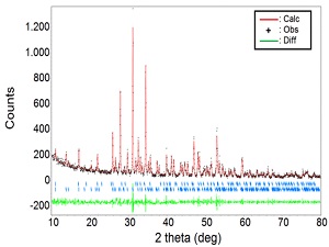
This study aimed to synthesize calcium phosphate loaded with silver nanoparticles and evaluate its antibacterial activity. We used cockle shells waste as raw material to prepare calcium phosphate. X-ray diffraction analysis showed that the constituent phases of calcium phosphate consist of hydroxyapatite (HA) and β-tricalcium phosphate (β-TCP). The incorporation process of silver nanoparticles on calcium phosphate was carried out in colloidal silver nanoparticles via the green-synthesis method using Citrus x microcarpa Bunge peel extract. The presence of colloidal silver nanoparticles through the green synthesis method was identified using UV-Vis spectrophotometer by the peak of the absorption band that occurred at 468 nm. The incorporation of silver nanoparticles into calcium phosphate did not significantly change the crystalline properties of HA and β-TCP. Evaluation of the antibacterial activity showed the silver nanoparticles had a strong antibacterial effect against Staphylococcus aureus, which also occurs in calcium phosphate loaded by silver nanoparticles. After being incorporated with silver nanoparticles, Calcium phosphate generally has no antibacterial effect. After being incorporated with silver nanoparticles, an inhibition zone with a diameter of about 9.8 mm can form. These results indicated that the method proposed in this study could be an alternative for developing calcium phosphate, which requires self-sterilization properties.
References
N. Eliaz and N. Metoki, “Calcium Phosphate Bioceramics: A Review of Their History, Structure, Properties, Coating Technologies and Biomedical Applications,” Materials (Basel)., vol. 10, no. 4, 2017, doi: 10.3390/ma10040334.
D. Arcos and M. Vallet-regí, “Europe PMC Funders Group Substituted Hydroxyapatite Coatings of Bone Implants,” vol. 8, no. 9, pp. 1781–1800, 2021, doi: 10.1039/C9TB02710F.
Y. Cao, L. Xiao, Y. Cao, A. Nanda, C. Xu, and Q. Ye, “3D Printed β-TCP Scaffold with Sphingosine 1-Phosphate Coating Promotes Osteogenesis and Inhibits Inflammation,” Biochem. Biophys. Res. Commun., vol. 512, no. 4, pp. 889–895, 2019, doi: 10.1016/j.bbrc.2019.03.132.
M. Taherimehr, R. Bagheri, and M. Taherimehr, “In-Vitro Evaluation of Thermoplastic Starch/Beta-Tricalcium Phosphate Nano-Biocomposite in Bone Tissue Engineering,” Ceram. Int., vol. 47, no. 11, pp. 15458–15463, 2021, doi: 10.1016/j.ceramint.2021.02.111.
L. Zhao et al., “Synthesis and Characterization of Silver-Incorporated Calcium Phosphate Antibacterial Nanocomposites for Mask Filtration Material,” Compos. Part B Eng., vol. 153, no. August, pp. 387–392, 2018, doi: 10.1016/j.compositesb.2018.09.004.
S. Mukherjee, S. Kumar, R. K. Sahu, and S. Nayar, “PVA-Graphene-Hydroxyapatite Electrospun Fibres as Air-Filters,” Mater. Res. Express, vol. 6, no. 12, p. 125366, Jan. 2020, doi: 10.1088/2053-1591/ab6ac9.
Y. Q. Shen, Y. J. Zhu, F. F. Chen, Y. Y. Jiang, Z. C. Xiong, and F. Chen, “Antibacterial Gluey Silver-Calcium Phosphate Composites for Dentine Remineralization,” J. Mater. Chem. B, vol. 6, no. 30, pp. 4985–4994, 2018, doi: 10.1039/C8TB00881G.
M. N. Capela et al., “Bioactivity and Antibacterial Activity Against E-Coli of Calcium-Phosphate-Based Glasses: Effect of Silver Content and Crystallinity,” Ceram. Int., vol. 43, no. 16, pp. 13800–13809, 2017, doi: 10.1016/j.ceramint.2017.07.100.
F. Afriani, E. J, Z. Zaitun, and Y. Tiandho, “Improvement of Hardness of Hydroxyapatite by the Addition of Silica from Tin Tailings,” J. Eng. Sci. Res., vol. 2, no. 2, pp. 85–89, 2020, doi: 10.23960/jesr.v2i2.48.
M. T. Islam, R. M. Felfel, E. A. Abou Neel, D. M. Grant, I. Ahmed, and K. M. Z. Hossain, “Bioactive Calcium Phosphate–Based Glasses and Ceramics and Their Biomedical Applications: A Review,” J. Tissue Eng., vol. 8, 2017, doi: 10.1177/2041731417719170.
F. Afriani, K. Dahlan, S. Nikmatin, and O. Zuas, “Alginate Affecting the Characteristics of Porous Beta-TCP/Alginate Composite Scaffolds,” J. Optoelectron. Biomed. Mater., vol. 7, no. 3, pp. 67–76, 2015, [Online]. Available: https://chalcogen.ro/67_Afriani.pdf.
D. G. Syarif, D. H. Prajitno, A. Kurniawan, M. B. Febrian, and R. Lesmana, “Hydrothermally Synthesis and Characterization of HAp and Zr-doped HAp Nanoparticles From Bovine Bone and Zircon for Photodynamic Therapy,” Process. Appl. Ceram., vol. 15, no. 2, pp. 146–153, 2021, doi: 10.2298/PAC2102146S.
M. R. Hasan, N. S. Mohd Yasin M. S. Mohd Ghazali, and N. F. Mohtar, “Proximate and Morphological Characteristics of Nano Hydroxyapatite (Nano Hap) Extracted From Fish Bone,” J. Sustain. Sci. Manag., vol. 15, no. 8, pp. 9–21, 2020, doi: 10.46754/jssm.2020.12.002.
F. Afriani, Siswoyo, R. Amelia, M. Hudatwi, Zaitun, and Y. Tiandho, “Hydroxyapatite From Natural Sources: Methods and Its Characteristics,” IOP Conf. Ser. Earth Environ. Sci., vol. 599, no. 1, pp. 0–7, 2020, doi: 10.1088/1755-1315/599/1/012055.
C. G. A. Das et al., “Antibacterial Activity of Silver Nanoparticles (Biosynthesis): A Short Review on Recent Advances,” Biocatal. Agric. Biotechnol., vol. 27, p. 101593, 2020, doi: 10.1016/j.bcab.2020.101593.
I. X. Yin, J. Zhang, I. S. Zhao, M. L. Mei, Q. Li, and C. H. Chu, “The Antibacterial Mechanism of Silver Nanoparticles and Its Application in Dentistry,” Int. J. Nanomedicine, vol. 15, pp. 2555–2562, 2020, doi: 10.2147/IJN.S246764.
A. M. Awwad, N. M. Salem, M. M. Aqarbeh, and F. M. Abdulaziz, “Green Synthesis, Characterization of Silver Sulfide Nanoparticles and Antibacterial Activity Evaluation,” Chem. Int., vol. 6, no. 1, pp. 42–48, 2020, doi: 10.5281/zenodo.3243157.
T. Ahmad, M. A. Bustam, M. Irfan, M. Moniruzzaman, H. M. A. Asghar, and S. Bhattacharjee, “Mechanistic Investigation of Phytochemicals Involved in Green Synthesis of Gold Nanoparticles Using Aqueous Elaeis Guineensis Leaves Extract: Role of Phenolic Compounds and Flavonoids,” Biotechnol. Appl. Biochem., vol. 66, no. 4, pp. 698–708, 2019, doi: 10.1002/bab.1787.
D. Bazin et al., “Diffraction Techniques and Vibrational Spectroscopy Opportunities to Characterise Bones,” Osteoporos. Int., vol. 20, no. 6, pp. 1065–1075, 2009, doi: 10.1007/s00198-009-0868-3.
B. Dickens, L. W. Schroeder, and W. E. Brown, “Crystallographic Studies of the Role of Mg as A Stabilizing Impurity in β-Ca3(PO4)2. The Crystal Structure of Pure β-Ca3(PO4)2,” J. Solid State Chem., vol. 10, no. 3, pp. 232–248, 1974, doi: 10.1016/0022-4596(74)90030-9.
F. Afriani, Mustari, and Y. Tiandho, “Pengaruh Lama Pemanasan terhadap Karakteristik Kristal Kalsium dari Limbah Cangkang Kerang [Effect of Heating Time on the Characteristics of Calcium Crystals from Shell Shell Waste],” J. EduMatSains, vol. 2, no. 2, pp. 189–200, 2018, doi: 10.33541/edumatsains.v2i2.606.
K.-R. Kang et al., “Synthesis and Characterization of β-Tricalcium Phosphate Derived From Haliotis sp. Shells,” Implant Dent., vol. 26, no. 3, pp. 378–387, Jun. 2017, doi: 10.1097/ID.0000000000000559.
S. Mohan, Y. Singh, D. K. Verma, and S. H. Hasan, “Synthesis of CuO Nanoparticles Through Green Route Using Citrus Limon Juice and Its Application as Nanosorbent for Cr(VI) remediation: Process Optimization with RSM and ANN-GA Based Model,” Process Saf. Environ. Prot., vol. 96, pp. 156–166, 2015, doi: 10.1016/j.psep.2015.05.005.
T. C. Prathna, N. Chandrasekaran, A. M. Raichur, and A. Mukherjee, “Biomimetic Synthesis of Silver Nanoparticles by Citrus Limon (Lemon) Aqueous Extract and Theoretical Prediction of Particle Size,” Colloids Surfaces B Biointerfaces, vol. 82, no. 1, pp. 152–159, 2011, doi: 10.1016/j.colsurfb.2010.08.036.
K. Momma and F. Izumi, “VESTA 3 for Three-Dimensional Visualization of Crystal, Volumetric and Morphology Data,” J. Appl. Crystallogr., vol. 44, no. 6, pp. 1272–1276, 2011, doi: 10.1107/S0021889811038970.
Downloads
Published
Issue
Section
License
Copyright (c) 2021 Yuant Tiandho, Rahmad Lingga, Evi.J Evi.J, Rifqi Almusawi Rafsanjani, Fitri Afriani

This work is licensed under a Creative Commons Attribution-ShareAlike 4.0 International License.
Authors who publish in IRiP agree to the following terms: Authors retain copyright and grant the IRiP right of first publication with the work simultaneously licensed under a Creative Commons Attribution License (CC BY-SA 4.0) that allows others to share (copy and redistribute the material in any medium or format) and adapt (remix, transform, and build upon the material) the work for any purpose, even commercially with an acknowledgment of the work's authorship and initial publication in IRiP. Authors are able to enter into separate, additional contractual arrangements for the non-exclusive distribution of the journal's published version of the work (e.g., post it to an institutional repository or publish it in a book), with an acknowledgment of its initial publication in IRiP. Authors are permitted and encouraged to post their work online (e.g., in institutional repositories or on their website) prior to and during the submission process, as it can lead to productive exchanges, as well as earlier and greater citation of published work (See The Effect of Open Access).