Identifying malaria disease through red-blood microscopic image with XGBoost and random forest methods
DOI:
https://doi.org/10.12928/bamme.v4i2.11740Keywords:
malaria, microscopic blood image, random forest, RShiny, XGBoostAbstract
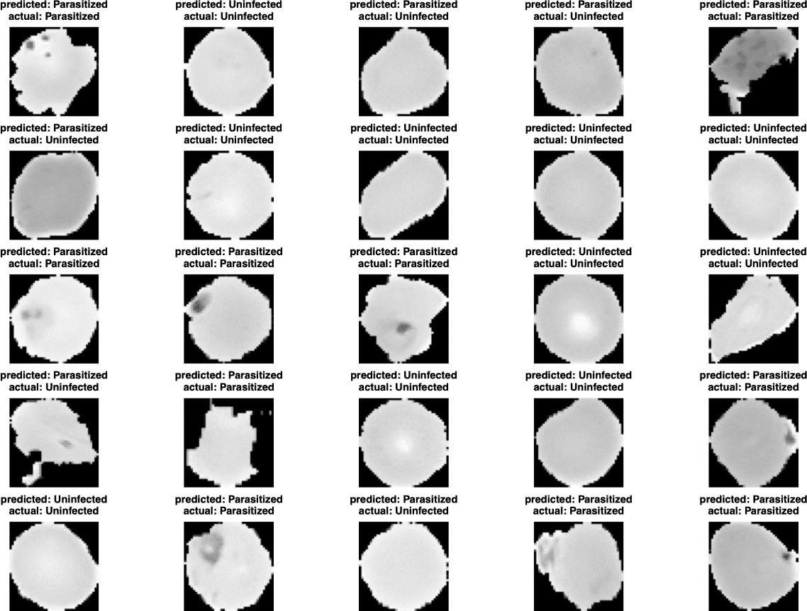
Blood cells that flow in the human body provide information to diagnose a disease. The information provided can be obtained through images of these blood cells using image processing techniques. Malaria is a very deadly disease and can affect everyone. Patients with malaria will experience anaemia because the red blood cells or erythrocytes are contaminated with plasmodium. This study offers an alternative solution to malaria disease identification through the image classification of red blood cells, by applying image processing and image classification methods with XGBoost and random forest. The research was conducted using the R programming language in RStudio and Python. The accuracy of XGBoost and random forest methods were 71.26% and 77.58%, respectively. Therefore, the random forest provided a better optimal classification model with higher accuracy. The model is used to build an application which is R web-based, RShiny. In practice, this application can be used by health workers in classifying patients based on red blood cell images such that the health centre would be easier to manage the existing patients.References
Bekele, A. (2017). Automatic detection of malaria parasite based on microscopic image analysis. Doctoral Dissertation. Addis Ababa University.
Berzosa, P., De Lucio, A., Romay-Barja, M., Herrador, Z., González, V., García, L., Fernández- Martínez, A., Santana-Morales, M., Ncogo, P., Valladares, B., Riloha, M., & Benito, A. (2018). Comparison of three diagnostic methods (microscopy, RDT, and PCR) for the detection of malaria parasites in representative samples from Equatorial Guinea. Malaria Journal, 17(1), 1–12. https://doi.org/10.1186/s12936-018-2481-4
Breiman, L. & Cutler, A. (2004). Random Forests. https://www.stat.berkeley.edu/~breiman/RandomForests/cc_home.htm
Breiman, L. & Cutler, A. (2003). Manual on setting up, using, and understanding random forest V4.0. https://www.stat.berkeley.edu/~breiman/Using_random_forests_v4.0.pdf
Breiman, L. (2001). Random Forest (45th ed.). Springer. https://link.springer.com/article/10.1023/A:1010933404324
Bridget, O. N. (2021). Machine-learning techniques for malaria incidence and tuberculosis prediction. Dissertation. African University of Science and Technology. http://repository.aust.edu.ng/xmlui/handle/123456789/5096
Chen, T., & Guestrin, C. (2016). XGBoost: A scalable tree boosting system. Proceedings of the ACM
SIGKDD International Conference on Knowledge Discovery and Data Mining, 13-17-Augu, 785– 794. https://doi.org/10.1145/2939672.2939785
Das, D. K., Ghosh, M., Pal, M., Maiti, A. K., & Chakraborty, C. (2013). Machine learning approach for automated screening of malaria parasite using light microscopic images. Micron, 45, 97–106. https://doi.org/10.1016/j.micron.2012.11.002
Dayat, A. R., & Banyal, N. A. (2018). Penyebab penyakit malaria dalam sel darah merah manusia dengan menggunakan support vektor machine (SVM) di Kota Jayapura-Papua. Jurnal ILKOM, 10(April), 28–32.
Endah, S. (2020). Mengenal malaria dan vektornya. Bandarlampung, 53(9).
Fitri, L. E., Widaningrum, T., Endharti, A. T., Prabowo, M. H., Winaris, N., & Nugraha, R. Y. B. (2022).
Malaria diagnostic update: From conventional to advanced method. Journal of Clinical Laboratory Analysis, 36(4), 1–14. https://doi.org/10.1002/jcla.24314
Fuhad, K. M. F., Tuba, J. F., Sarker, M. R. A., Momen, S., Mohammed, N., & Rahman, T. (2020). Detection from blood smear and its smartphone based application. Diagnostics, 10(329).
Gitta, B., & Kilian, N. (2020). Diagnosis of malaria parasites Plasmodium sp. in endemic areas:
Current strategies for an ancient disease. BioEssays, 42(1), 1–12. https://doi.org/10.1002/bies.201900138
Jain, N., Chauhan, A., Tripathi, P., Moosa, S. Bin, Aggarwal, P., & Oznacar, B. (2020). Cell image
analysis for malaria detection using deep convolutional network. Intelligent Decision Technologies, 14(1), 55–65. https://doi.org/10.3233/IDT-190079
Janitza, S., & Hornung, R. (2018). On the overestimation of random forest’s out-of-bag error. Plos One, 13(8). https://doi.org/10.1371/journal.pone.0201904
Jin, Z., Shang, J., Zhu, Q., Ling, C., Xie, W., & Qiang, B. (2020). RFRSF: Employee turnover prediction based on random forests and survival analysis. Lecture Notes in Computer Science (Including Subseries Lecture Notes in Artificial Intelligence and Lecture Notes in Bioinformatics), 12343 LNCS, 503–515. https://doi.org/10.1007/978-3-030-62008-0_35
Kapwata, T., & Gebreslasie, M. T. (2016). Random forest variable selection in spatial malaria transmission modelling in Mpumalanga Province South Africa. Geospatial Health, 11(3), 251– 262. https://doi.org/10.4081/gh.2016.434
Kementerian Kesehatan. (2017). Pedoman Teknis Pemeriksaan Parasit Malaria. Buku Pedoman, 1–78.
Khoirunnisa, A., & Ramadhan, N. G. (2023). Improving malaria prediction with ensemble learning and robust scaler: An integrated approach for enhanced accuracy. Jurnal Infotel, 15(4), 326– 334. https://doi.org/10.20895/infotel.v15i4.1056
Madabhushi, A., & Lee, G. (2016). Image analysis and machine learning in digital pathology: Challenges and opportunities. Medical Image Analysis, 33, 170–175. https://doi.org/10.1016/j.media.2016.06.037
Pan, W. D., Dong, Y., & Wu, D. (2018). Classification of malaria-infected cells using deep convolutional neural networks. Machine Learning - Advanced Techniques and Emerging Applications. https://doi.org/10.5772/intechopen.72426
PDPERSI. (2019). Buku Saku Penatalaksanaan Kasus Malaria.
http://www.pdpersi.co.id/kanalpersi/data/elibrary/bukusaku_malaria.pdf
Rahman, I. F. (2020). Implementasi Metode SVM, MLP dan Xgboost pada Data Ekspresi Gen. 1–79.
https://dspace.uii.ac.id/handle/123456789/23679
Rajaraman, S., Antani, S. K., Poostchi, M., Silamut, K., Hossain, M. A., Maude, R. J., Jaeger, S., & Thoma, G. R. (2018). Pre-trained convolutional neural networks as feature extractors toward improved malaria parasite detection in thin blood smear images. PeerJ, 2018(4), 1–17. https://doi.org/10.7717/peerj.4568
Segal, M. R. (2004). Machine Learning Benchmarks and Random Forest. Center for Bioinformatics and Molecular Biostatistics, 15. https://escholarship.org/uc/item/35x3v9t4
Shafila, G. A. (2020). Implementasi metode extreme gradient boosting (XGBoost) untuk klasifikasi pada data bioinformatika. Studi Kasus Penyakit Ebola, GSE 122692, 1– 77.
Tang, G. H., Rabie, A. B. M., & Hägg, U. (2004). Indian hedgehog: A mechanotransduction mediator in condylar cartilage. Journal of Dental Research, 83(5), 434–438. https://doi.org/10.1177/154405910408300516
Ummah, M. S. (2019). Properties of AdeABC and AdeIJK efflux systems of Acinetobacter baumannii compared with those of the AcrAB-TolC system of Escherichia coli. Sustainability (Switzerland), 11(1), 1–14.
WHO. (2023). World Malaria Report. https://www.who.int/teams/global-malaria-programme/reports/world-malaria-report-2023
WHO. (2018). World Malaria Report 2018. https://www.who.int/teams/global-malaria-programme/reports/world-malaria-report-2018
Yang, F., Poostchi, M., Yu, H., Zhou, Z., Silamut, K., Yu, J., Maude, R. J., Jaeger, S., & Antani, S. (2020). Deep Learning for Smartphone-Based Malaria Parasite Detection in Thick Blood Smears. IEEE Journal of Biomedical and Health Informatics, 24(5), 1427–1438. https://doi.org/10.1109/JBHI.2019.2939121

Downloads
Published
Issue
Section
License
Copyright (c) 2024 Rohmatul Fajriyah, Muhammad Muhajir, Ahmad Hussain Abdullah, Devina Gilar Ayu, Iqbal Fathur Rahma

This work is licensed under a Creative Commons Attribution-ShareAlike 4.0 International License.
Authors who publish with this journal agree to the following terms:
- Authors retain copyright and grant the journal right of first publication with the work simultaneously licensed under Creative Commons Attribution License that allows others to share the work with an acknowledgement of the work's authorship and initial publication in this journal.
- Authors are able to enter into separate, additional contractual arrangements for the non-exclusive distribution of the journal's published version of the work (e.g., post it to an institutional repository or publish it in a book), with an acknowledgement of its initial publication in this journal.
- Authors are permitted and encouraged to post their work online (e.g., in institutional repositories or on their website) prior to and during the submission process, as it can lead to productive exchanges, as well as earlier and greater citation of published work (See The Effect of Open Access).


